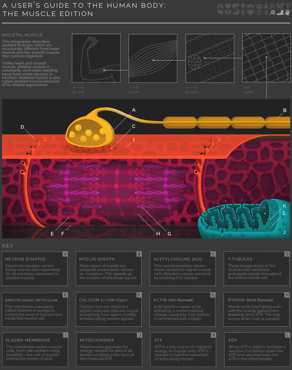
A muscle is basically a bundle of fibers wrapped in a connective tissue. Each fiber is made of a single, elongated muscle cell, and each cell is composed of myofibrils, which in turn are made of myofilaments. The thick myofilaments are made of myosin, and thin myofilament of Actin, Troponin, and Tropomyosin.
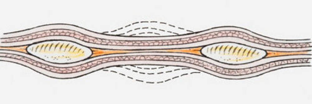
The primary mode of action for muscle is by contraction. When the CNS sends a signal, the thick and thin myosin filaments form a "crossbridge" pattern by sliding past each other. This makes the sarcomeres shorter and thicker, contracting the muscle.
- A signal is sent from the brain or the spinal cord to the muscle via neurons
- An action potential is generated in the neuron, releasing Ca++ in the neuromuscular junction
- The influx of caalcium ions causes acetylcholine (AcH) to be released in the synaptic cleft
- AcH binds to the AcH receptors present in the sarcolemma, increasing its permeability
- Na++enter the sarcolemma, changing its polarity, and creating an action potential
- Ca++ are released by the sarcoplasmic reticulum, as the action potential travels down the T-tubules in the muscle fiber
As the myosin head swivels, another ATP molecule binds to myosin, breaking the actin-myosin bridge. The ATP is again hydrolyzed, and last four steps of the process are repeated, making the sarcomere shorter and shorter, until adequate Ca++ and ATP are present. Many myosin heads move in the same direction and a number of times, to contract a single muscle.
When the nervous impulse stops, the calcium gates close, and the sarcoplasmic reticulum is no longer permeable. The Ca++ return to the sarcoplasmic reticulum, and troponin and tropomyosin are reverted to their original positions. With the binding sites blocked, myosin cannot form cross-bridges with actin, and the muscle relaxes.
Here is a list of few structures, to help you have a better understanding of the process -
- Ca++ bind with troponin C, causing the tropomyosin to shift, and expose the myosin binding sites on actin
- ATP is hydrolyzed into ADP and phosphorus, releasing energy for myosin power stroke
- Myosin binds to actin
- Myosin head bends and actin slides over the myosin surface
- Myosin releases the ADP molecule
As the myosin head swivels, another ATP molecule binds to myosin, breaking the actin-myosin bridge. The ATP is again hydrolyzed, and last four steps of the process are repeated, making the sarcomere shorter and shorter, until adequate Ca++ and ATP are present. Many myosin heads move in the same direction and a number of times, to contract a single muscle.
When the nervous impulse stops, the calcium gates close, and the sarcoplasmic reticulum is no longer permeable. The Ca++ return to the sarcoplasmic reticulum, and troponin and tropomyosin are reverted to their original positions. With the binding sites blocked, myosin cannot form cross-bridges with actin, and the muscle relaxes.
Here is a list of few structures, to help you have a better understanding of the process -
- Myofibrils - Thin fibers in the muscle cells
- Sarcomere - A structural unit of myofibril
- Sarcoplasmic Reticulum - Tubules surrounding myofibrils responsible for storing and diffusing Ca ions
- Sarcolemma - Cell membrane of muscle cells
- T-tubules - Tubules running through the muscle fibers through which Ca++ flow
- Troponin - A complex of proteins, which combine with Calcium ions, and shift tropomyosin
- Tropomyosin - Protein component of muscle fiber, which in its natural state, blocks myosin-actin binding sites
